
Home
Common Techniques
Classroom Experiments
Virtual Experiments
Tutorials
Games
Glossary
Links
Publishing
Opportunities
About This Site
Contact Us
ZFIN
Cite Us
Developmental Staging Experiment
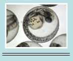
This picture depicts a 16 cell embryo in the clevage period. You can see three different layers of cells in the picture (denoted by blue circles).
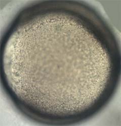
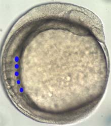
Both of the pictures are of a cell in 80% epiboly, which is in the blastula period. The blue circles in the vegetal view picture depict the boundary between the cells and yolk.
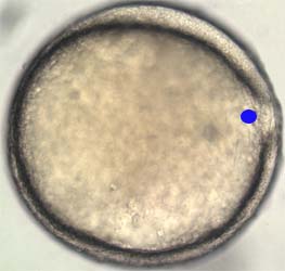
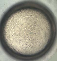
The embryo above is in 95% epiboly, which is also in the blastula period. The 5% of yolk that isn't covered is the light area to the right of the blue circle.
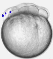
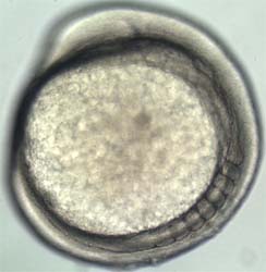
The embryo above is in the segmentation period. This particular embryo has 5 somites. The blue circles are located to the right of the somites.
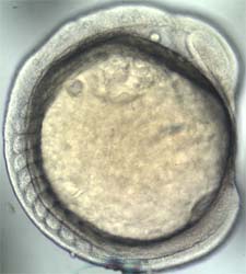
The embryo above is also in the segmentation period. If you count, you'll notice that the embryo has 7 somites.
The embryo is in the segmentation period. This particular embryo has 10 somites.
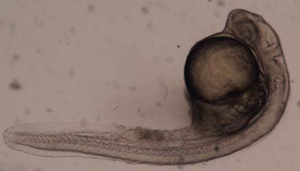
This embryo is one day old and is in the pharyngula period.
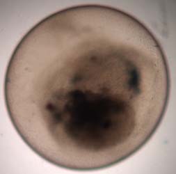
This embry is dead. The dead cells are the reason for the black color within the chrion.